

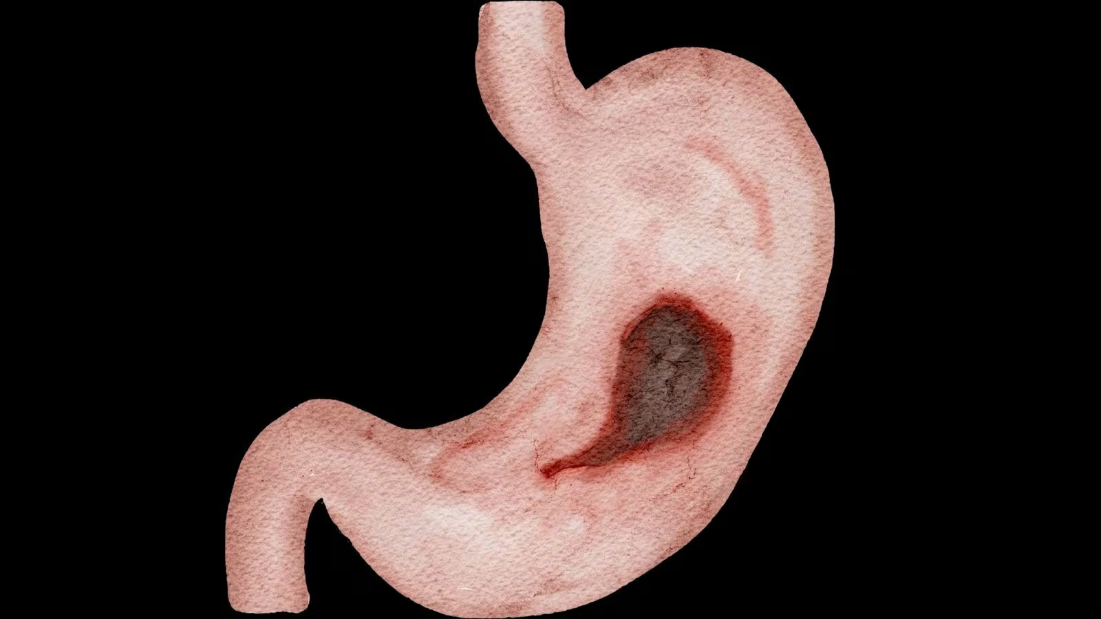

A recent scientific investigation conducted in Brazil has introduced a promising approach to improving the detection of gastric cancer by examining a biological material that has historically been overlooked. The study, led by researchers at a leading oncology centre and published in a peer-reviewed journal, highlights the diagnostic value of gastric fluid collected during routine endoscopic procedures.
In clinical practice, upper gastrointestinal endoscopy is widely used to evaluate the stomach and identify abnormalities. During the procedure, gastric fluid is often collected to facilitate visualisation but is typically discarded without further analysis. However, researchers in Brazil have demonstrated that this fluid may contain important molecular indicators associated with the presence of cancer.
The study focused on the detection of cell-free DNA within gastric fluid. This genetic material is released into bodily fluids when cells undergo stress, damage or death. In the context of cancer, these processes tend to occur more frequently due to rapid tumour growth, increased cellular turnover and inflammatory responses within the affected tissue.
As a result, gastric fluid can accumulate fragments of DNA that originate from different regions of the stomach. According to the findings, analysing these fragments may provide a broader representation of tissue alterations compared to traditional biopsy methods, which rely on small, localised samples.
Biopsy remains the gold standard for confirming gastric cancer; however, it has recognised limitations. In certain cases, lesions may be difficult to access, or tumour cells may be distributed unevenly, reducing the likelihood of accurate sampling. This is particularly relevant in early-stage or diffuse forms of the disease, where subtle changes may not be captured in a single tissue specimen.
The Brazilian study suggests that incorporating gastric fluid analysis into existing diagnostic protocols could complement biopsy findings, potentially increasing sensitivity in detecting malignancies. Importantly, this approach does not require additional invasive procedures, as the material is already obtained during routine examinations. Consequently, it does not increase procedural time, cost or patient discomfort.
Another notable observation from the research relates to the potential prognostic value of cell-free DNA levels. In some cases, higher concentrations of DNA fragments were associated with more favourable clinical outcomes. Researchers propose that this may reflect a more active immune response, indicating that the body is mounting a stronger defence against tumour cells.
Despite these encouraging findings, the authors emphasise that the method is not yet suitable for standalone diagnostic use. Elevated levels of cell-free DNA can also be present in non-malignant conditions, such as gastritis or other inflammatory processes, which may limit its specificity.
Further research is required to validate these results across larger and more diverse populations, as well as to refine the interpretation of molecular markers identified in gastric fluid. Scientists also highlight the need to better understand the biological origin of the detected DNA and its relationship with tumour behaviour.
The study represents an important contribution from Brazil to the global effort to improve cancer diagnostics. By re-evaluating materials already available in routine medical procedures, researchers are opening new pathways for more comprehensive, accessible and less invasive approaches to disease detection.
As advances in molecular diagnostics continue, such strategies may play a significant role in enhancing early detection and improving outcomes for patients with gastric cancer worldwide.